|
April 2005
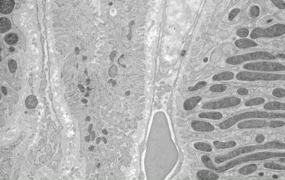
Mouse Kidney prepared by high pressure freezing and freeze substitution. Micrograph courtesy of Richard M. Hays MD, Leslie
Gunther-Cummins and Frank Macaluso, Albert Einstein College of Medicine
March 2005
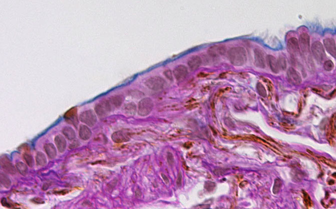
Human lung tissue stained with Periodic Acid Schiff (PAS), Alcian blue, and hematoxylin, with immunoperoxidase staining
for surfactant protein. Contributed by Barbara Ferris and Philip Leopold, Department of Genetic Medicine, Weill Medical College
of Cornell University.
February 2005
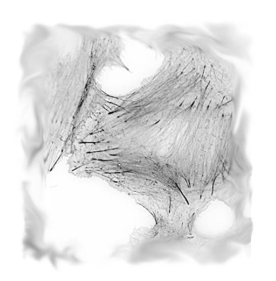
Oregon green-phalloidin stain of actin microfilaments in an A549 lung epithelial cell. The image was acquired as a grayscale
image of the green channel under epifluorescence illumination. In the final image, the contrast was inverted and the
edges of the image were dodged in Photoshop. Contributed by Philip Leopold, Department of Genetic Medicine, Weill Medical
College of Cornell University.
|